خلية محببة
الخلية المُحَبَّبَة[2] (بالإنجليزية: Granulocyte) أحد التقسيمات الرئيسية للخلايا البيض وهي تشكل معظمها بنسبة 50-70%.[3][4][5] تأتي من خط الخلايا الجذعية كباقي مكونات الدم، وتمتاز بأنها كرية الشكل غير مقسمة وقصيرة الحياة حوالي 2-5 أيام ثم تتجدد. تستطيع أن تتدخل في الأنسجة، وذلك مخترقة الأوعية الدموية والغشاء الخلوي. وتضم هذه المجموعة 3 أنواع من الخلايا البيض:
| خلية محببة | |
|---|---|
| تفاصيل | |
| نوع من | خلية دم بيضاء[1] |
| FMA | 62854 |
| ن.ف.م.ط. | A11.118.637.415، وA11.148.350، وA11.627.340، وA15.145.229.637.415، وA15.378.316.340، وA15.382.490.315 |
| ن.ف.م.ط. | D006098 |
| تعديل مصدري - تعديل | |

سميت بالخلايا المحببة نظرا إلى احتوائها على فقاقيع تحوي سائل مقاوم للميكروبات. بهذ تنتمي إلى النظام المناعي في الجسم. تصنيف الثلاثة أنواع منها يعتمد على نوع الصبغة التي تتقبلها الخلية، فمثلا الخلية العدلة تأخذ الصبغتين الحامضية والقاعدية عند الكشف عنهم.
منشأها
عدلتتكون الخلايا المحببة في نخاع العظام، وسميت بهذا الاسم لوجود حبيبات صغيرة في سائل السايتوبلازم.
بعض أشكالها:
-
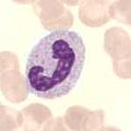 عصوي
عصوي -
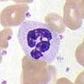 مجزأ
مجزأ -
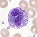 حبيبي
حبيبي -


انظر أيضا
عدلمراجع
عدل- ^ نموذج تأسيسي في التشريح، QID:Q1406710
- ^ المعجم الطبي الموحد
- ^ Polyzoidis S، Koletsa T، Panagiotidou S، Ashkan K، Theoharides TC (2015). "Mast cells in meningiomas and brain inflammation". J Neuroinflammation. ج. 12 ع. 1: 170. DOI:10.1186/s12974-015-0388-3. PMC:4573939. PMID:26377554.
MCs originate from a bone marrow progenitor and subsequently develop different phenotype characteristics locally in tissues. Their range of functions is wide and includes participation in allergic reactions, innate and adaptive immunity, inflammation, and autoimmunity [34]. In the human brain, MCs can be located in various areas, such as the pituitary stalk, the pineal gland, the area postrema, the choroid plexus, thalamus, hypothalamus, and the median eminence [35]. In the meninges, they are found within the dural layer in association with vessels and terminals of meningeal nociceptors [36]. MCs have a distinct feature compared to other hematopoietic cells in that they reside in the brain [37]. MCs contain numerous granules and secrete an abundance of prestored mediators such as corticotropin-releasing hormone (CRH), neurotensin (NT), substance P (SP), tryptase, chymase, vasoactive intestinal peptide (VIP), vascular endothelial growth factor (VEGF), TNF, prostaglandins, leukotrienes, and varieties of chemokines and cytokines some of which are known to disrupt the integrity of the blood-brain barrier (BBB) [38–40].
They key role of MCs in inflammation [34] and in the disruption of the BBB [41–43] suggests areas of importance for novel therapy research. Increasing evidence also indicates that MCs participate in neuroinflammation directly [44–46] and through microglia stimulation [47], contributing to the pathogenesis of such conditions such as headaches, [48] autism [49], and chronic fatigue syndrome [50]. In fact, a recent review indicated that peripheral inflammatory stimuli can cause microglia activation [51], thus possibly involving MCs outside the brain.{{استشهاد بدورية محكمة}}: صيانة الاستشهاد: دوي مجاني غير معلم (link) - ^ Mayer، Gene (2006). "Immunology — Chapter One: Innate (non-specific) Immunity". Microbiology and Immunology On-Line Textbook. USC School of Medicine. مؤرشف من الأصل في 2014-10-21. اطلع عليه بتاريخ 2008-11-12.
- ^ Lee DM، Friend DS، Gurish MF، Benoist C، Mathis D، Brenner MB (سبتمبر 2002). "Mast cells: a cellular link between autoantibodies and inflammatory arthritis". Science. ج. 297 ع. 5587: 1689–92. DOI:10.1126/science.1073176. PMID:12215644.
| خلية محببة في المشاريع الشقيقة: | |
| |
هذه بذرة مقالة عن علم الأحياء الخلوي بحاجة للتوسيع. فضلًا شارك في تحريرها. |