ملف:Variant Creutzfeldt-Jakob disease (vCJD), H&E.jpg
لا توجد دقة أعلى متوفرة.
Variant_Creutzfeldt-Jakob_disease_(vCJD),_H&E.jpg (700 × 554 بكسل حجم الملف: 80 كيلوبايت، نوع MIME: image/jpeg)
| هذا ملف من ويكيميديا كومنز. معلومات من صفحة وصفه مبينة في الأسفل. كومنز مستودع ملفات ميديا ذو رخصة حرة. |
,_H%26E.jpg)
ملخص
| الوصف |
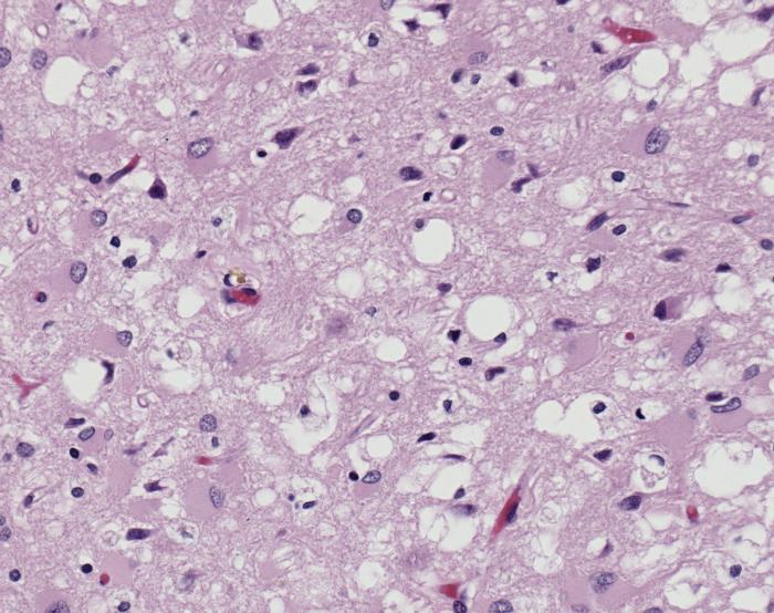
ID#: 10131 Magnified 100X, and stained with H&E (hematoxylin and eosin) staining technique, this light photomicrograph of brain tissue reveals the presence of prominent spongiotic changes in the cortex, and loss of neurons in a case of variant Creutzfeldt-Jakob disease (vCJD). Variant Creutzfeldt-Jakob disease (vCJD) is a prion disease that was first described in 1996 in the United Kingdom. There is now strong scientific evidence that the agent responsible for the outbreak of prion disease in cows, bovine spongiform encephalopathy (BSE or 'mad cow' disease), is the same agent responsible for the outbreak of vCJD in humans. Both disorders are invariably fatal brain diseases with unusually long incubation periods measured in years, and are caused by an unconventional transmissible agent called a prion. vCJD is not the same disease as classic CJD. It has different clinical and pathologic characteristics from classic CJD. Each disease also has a particular genetic profile of the prion protein gene. |
| المصدر | Public Health Image Library (PHIL) ID#: 10131 |
| المؤلف |
Content Providers(s): CDC/ Teresa Hammett Photo Credit: Sherif Zaki; MD; PhD; Wun-Ju Shieh; MD; PhD; MPH |
| الترخيص (إعادة استخدام هذا الملف) |
Copyright Restrictions: None - This image is in the public domain and thus free of any copyright restrictions. As a matter of courtesy we request that the content provider be credited and notified in any public or private usage of this image. |
ترخيص
هذه الصُّورة مِن إِنتاج مراكز السيطرة على الأمراض والوقاية منها (CDC)، التَّابِعة لوزارة الصِّحة والخدمات البشريَّة الأمريكيَّة، التقطت أو أُنشئت بصفتها جزءاً مِن عمل المُوظَفين الرَّسميين فيها. ولأَنَّها عملٌ من إِنتاج الحكومة الاتحادية للولايات المُتحدة الأمريكيَّة، فإِنَّ هذه الصُّورة في النِّطاق العامِّ.
|
تاريخ الملف
اضغط على زمن/تاريخ لرؤية الملف كما بدا في هذا الزمن.
| زمن/تاريخ | صورة مصغرة | الأبعاد | مستخدم | تعليق | |
|---|---|---|---|---|---|
| حالي | 19:55، 30 يناير 2008 | | 700 × 554 (80 كيلوبايت) | Patho | {{Information| |Description=ID#: 10131 Magnified 100X, and stained with H&E (hematoxylin and eosin) staining technique, this light photomicrograph of brain tissue reveals the presence of prominent spongiotic changes in the cortex, and loss of neurons in |
استخدام الملف
الصفحة التالية تستخدم هذا الملف:
الاستخدام العالمي للملف
الويكيات الأخرى التالية تستخدم هذا الملف:
- الاستخدام في de.wikibooks.org
- الاستخدام في en.wikipedia.org
- الاستخدام في es.wikipedia.org
- الاستخدام في sr.wikipedia.org
,_H%26E.jpg)