ملف:U fibres big.JPG

الملف الأصلي (1٬638 × 1٬458 بكسل حجم الملف: 185 كيلوبايت، نوع MIME: image/jpeg)
| هذا ملف من ويكيميديا كومنز. معلومات من صفحة وصفه مبينة في الأسفل. كومنز مستودع ملفات ميديا ذو رخصة حرة. |
ملخص
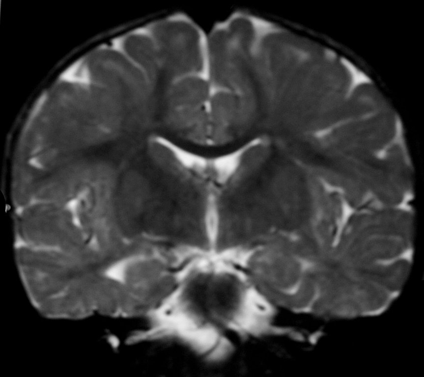
| الوصف | Desmyelinating Disorder |
| التاريخ | |
| المصدر | Radiology picture of the day |
| المؤلف | Dr. Laughlin Dawes |
| الترخيص (إعادة استخدام هذا الملف) |
Explanation
- "This 18-month old female presented with developmental delay. By this age myelination should be essentially
complete, yet there is residual T2 high signal in the subcortical white matter due to disruption of normal myelin formation. The differential diagnosis is depends on the head size.
With megalencephaly the possibilities are:
- Alexander's disease
- Canavan's disease
- van der Knapp’s leukodystrophy (but there is no cystic change)
Without megalencephaly: - in utero infection including Toxoplasma, Rubella, cytomegalovirus and herpes simplex virus. - mitochondrial cytopathy - Pelizeus-Merzbacher disease (but patient is female and PMD is X-linked recessive) - 18q deletion syndrome (the karyotype was normal) - Tuberous sclerosis (there were no other stigmata)
The more common leukodystrophies, metachromatic leukodystrophy and adrenoleukodystrophy, typically spare the subcortical U-fibres.
Credit: Dr Laughlin Dawes"Esta niña de 18 meses presenta retraso en el desarrollo. A esta edad la mielinización debería, en esencia, estar completa, aunque aún hay una alta señal de T2 en la materia blanca subcortical debido a la disrupción de la formación normal de mielina. El diagnóstico diferencial depende del volumen cefálico:
Con macrocefalia las posibilidades son:
- Enfermedad de alexander
- Enfermedad de Canavan
- Leucodistrofia de van der Knaap (sin cambios císticos)
Sin macrocefalia:
- Infecciones intrauterinas como toxoplasmosis, rubella, citomegalovirus y virus herpes simplex.
- Citopatía mitocondrial
- Enfermedad de Pelizeus-Merzbacher (Pero el paciente es niña y la enfermedad está ligada al X de forma recesiva)
- Sindrome de deleción del cromosoma 18q (El cariotipo era normal)
- Esclerosis tuberosa (no habia otras llagas)
Autor Dr Laughlin Dawes
ترخيص
- يحقُّ لك:
- مشاركة العمل – نسخ العمل وتوزيعه وبثُّه
- إعادة إنتاج العمل – تعديل العمل
- حسب الشروط التالية:
- نسب العمل إلى مُؤَلِّفه – يلزم نسب العمل إلى مُؤَلِّفه بشكل مناسب وتوفير رابط للرخصة وتحديد ما إذا أجريت تغييرات. بالإمكان القيام بذلك بأية طريقة معقولة، ولكن ليس بأية طريقة تشير إلى أن المرخِّص يوافقك على الاستعمال.
تاريخ الملف
اضغط على زمن/تاريخ لرؤية الملف كما بدا في هذا الزمن.
| زمن/تاريخ | صورة مصغرة | الأبعاد | مستخدم | تعليق | |
|---|---|---|---|---|---|
| حالي | 16:49، 10 مارس 2008 | | 1٬638 × 1٬458 (185 كيلوبايت) | Gustavocarra | {{Information |Description=Desmyelinating Disorder |Source=[http://www.radpod.org/2006/11/19/dysmyelinating-disorder/ Radiology picture of the day] |Date=19/11/2006 |Author=Dr. Laughlin Dawes |Permission=Creative Commons |other_versions= }} ==Explanation |
استخدام الملف
الصفحة التالية تستخدم هذا الملف:
الاستخدام العالمي للملف
الويكيات الأخرى التالية تستخدم هذا الملف:
- الاستخدام في de.wikipedia.org
- الاستخدام في en.wikipedia.org
- الاستخدام في es.wikipedia.org
- الاستخدام في it.wikipedia.org
- الاستخدام في ru.wikipedia.org
- الاستخدام في sr.wikipedia.org