ملف:Electron micrograph of neuromuscular junction (cross-section).jpg
لا توجد دقة أعلى متوفرة.
Electron_micrograph_of_neuromuscular_junction_(cross-section).jpg (433 × 289 بكسل حجم الملف: 95 كيلوبايت، نوع MIME: image/jpeg)
| هذا ملف من ويكيميديا كومنز. معلومات من صفحة وصفه مبينة في الأسفل. كومنز مستودع ملفات ميديا ذو رخصة حرة. |
.jpg)
ملخص
| الوصف |
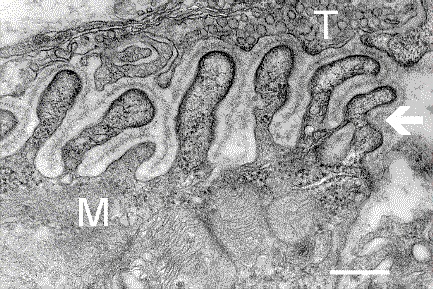
English: Electron micrograph showing a cross-section through the neuromuscular junction. T is the axon terminal, M is the muscle fiber. The arrow shows junctional folds with basal lamina. Postsynaptic densities are visible on the tips between the folds. The scale is 0.3 µm. |
| التاريخ | Originally uploaded to en.wikipedia on ١٠ مارس ٢٠٠٦. |
| المصدر | Synapse Web at the National Institute of Mental Health, National Institutes of Health; originally from en.wikipedia; description page is/was here. |
| المؤلف | National Institute of Mental Health; originally uploaded by Nrets at en.wikipedia. |
ترخيص
هذه الصُّورة هي عملٌ مِن إِنتاج معاهد الصِّحة الوطنية الأَمريكيَّة، وهي جزءٌ من وزارة الصِّحة والخدمات البشريَّة في الولايات المُتحدة الأمريكيَّة. وبما أنَّها عملٌ من إِنتاج الحكومة الاتحادية للولايات المُتحدة، فإِنَّ هذه الصُّورة في تقعُ في النِّطاق العامّ.
 |
||
| هذا الملفُّ مَلحُوظُ بصفته غيرَ مُقيَّدٍ بحقوق التَّأليف والنشر، وهذا يشمل أيضاً الحقوق المُجاوِرة أَو ذات الصلة جميعُها. | ||
سجلُّ الرَّفع الأصيل
(All user names refer to en.wikipedia)
- 2006-03-10 20:07 Nrets 433×289×8 (97758 bytes) Electron micrograph showing a cross section through the neuromuscular junction. T is the axon terminal, M is the muscle fiber. The arrow shows junctional folds with basal lamina. Postsynaptic densities are visible on the tips between the folds. Scale is 0
تاريخ الملف
اضغط على زمن/تاريخ لرؤية الملف كما بدا في هذا الزمن.
| زمن/تاريخ | صورة مصغرة | الأبعاد | مستخدم | تعليق | |
|---|---|---|---|---|---|
| حالي | 03:41، 22 مارس 2007 | | 433 × 289 (95 كيلوبايت) | Fran Rogers | {{Information |Description=Electron micrograph showing a cross section through the neuromuscular junction. T is the axon terminal, M is the muscle fiber. The arrow shows junctional folds with basal lamina. Postsynaptic densities are visible on the tips be |
استخدام الملف
الصفحة التالية تستخدم هذا الملف:
الاستخدام العالمي للملف
الويكيات الأخرى التالية تستخدم هذا الملف:
- الاستخدام في cs.wikipedia.org
- الاستخدام في de.wikipedia.org
- الاستخدام في en.wikipedia.org
- الاستخدام في es.wikipedia.org
- الاستخدام في fa.wikipedia.org
- الاستخدام في gl.wikipedia.org
- الاستخدام في he.wikipedia.org
- الاستخدام في ko.wikipedia.org
- الاستخدام في pt.wikipedia.org
- الاستخدام في ru.wikipedia.org
- الاستخدام في uk.wikipedia.org
- الاستخدام في zh.wikipedia.org
.jpg)