ملف:Carboxysome 3 images.png

حجم هذه المعاينة: 800 × 369 بكسل. الأبعاد الأخرى: 320 × 147 بكسل | 640 × 295 بكسل | 1٬024 × 472 بكسل | 1٬280 × 590 بكسل | 2٬943 × 1٬356 بكسل.
الملف الأصلي (2٬943 × 1٬356 بكسل حجم الملف: 3٫72 ميجابايت، نوع MIME: image/png)
| هذا ملف من ويكيميديا كومنز. معلومات من صفحة وصفه مبينة في الأسفل. كومنز مستودع ملفات ميديا ذو رخصة حرة. |
ملخص
| الوصف |
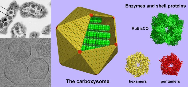
English: (Left, above) A thin-section electron micrograph of H. neapolitanus cells with carboxysomes inside. In one of the cells shown, arrows highlight the visible carboxysomes. (Left, below) Purified carboxysomes (material courtesy of S. Heinhorst and G. Cannon) as visualized by cryo-electron microscopy (courtesy of M. Yeager and K. Dryden). (right) Models for the structure of the carboxysome. Current data suggest that the shell is composed of several hundred hexameric protein building blocks and 12 pentameric building blocks. The three-dimensional atomic structures of the shell proteins have been determined by X-ray crystallography. RuBisCO, the main interior enzyme is shown packed inside in a regular arrangement for simplicity, though the actual organization of the enzymes is not understood yet. The other key enzyme, carbonic anhydrase, which is present in lesser amounts, is not illustrated. Scale bars are 100 nm. (image by T. Yeates). |
| المصدر | Modified version of Image:Carboxysome.png incorporating image taken from Image:Carboxysomes EM.jpg |
| المؤلف | Prof. Todd O. Yeates, UCLA Dept. of Chem. and Biochem. |
| إصدارات أخرى |
[]
PNG:
|


ترخيص
هذا الملف مُرخص تحت رخصة المشاع المبدع نسبة المصنف إلى مؤلفه 3.0 العامة
- يحقُّ لك:
- مشاركة العمل – نسخ العمل وتوزيعه وبثُّه
- إعادة إنتاج العمل – تعديل العمل
- حسب الشروط التالية:
- نسب العمل إلى مُؤَلِّفه – يلزم نسب العمل إلى مُؤَلِّفه بشكل مناسب وتوفير رابط للرخصة وتحديد ما إذا أجريت تغييرات. بالإمكان القيام بذلك بأية طريقة معقولة، ولكن ليس بأية طريقة تشير إلى أن المرخِّص يوافقك على الاستعمال.
تاريخ الملف
اضغط على زمن/تاريخ لرؤية الملف كما بدا في هذا الزمن.
| زمن/تاريخ | صورة مصغرة | الأبعاد | مستخدم | تعليق | |
|---|---|---|---|---|---|
| حالي | 01:38، 6 أغسطس 2008 | | 2٬943 × 1٬356 (3٫72 ميجابايت) | TimVickers | {{Information |Description={{en|1=(Left, above) A thin-section electron micrograph of H. neapolitanus cells with carboxysomes inside. In one of the cells shown, arrows highlight the visible carboxysomes. (Left, below) Purified carboxysomes (material court |
استخدام الملف
الصفحة التالية تستخدم هذا الملف:
الاستخدام العالمي للملف
الويكيات الأخرى التالية تستخدم هذا الملف:
- الاستخدام في bg.wikipedia.org
- الاستخدام في en.wikipedia.org
- الاستخدام في en.wikiversity.org
- الاستخدام في sw.wikipedia.org
- الاستخدام في tr.wikipedia.org