ملف:Hippel Lindau.gif

حجم هذه المعاينة: 508 × 599 بكسل. الأبعاد الأخرى: 204 × 240 بكسل | 407 × 480 بكسل | 805 × 949 بكسل.
الملف الأصلي (805 × 949 بكسل حجم الملف: 278 كيلوبايت، نوع MIME: image/gif)
| هذا ملف من ويكيميديا كومنز. معلومات من صفحة وصفه مبينة في الأسفل. كومنز مستودع ملفات ميديا ذو رخصة حرة. |
ملخص
| الوصف |
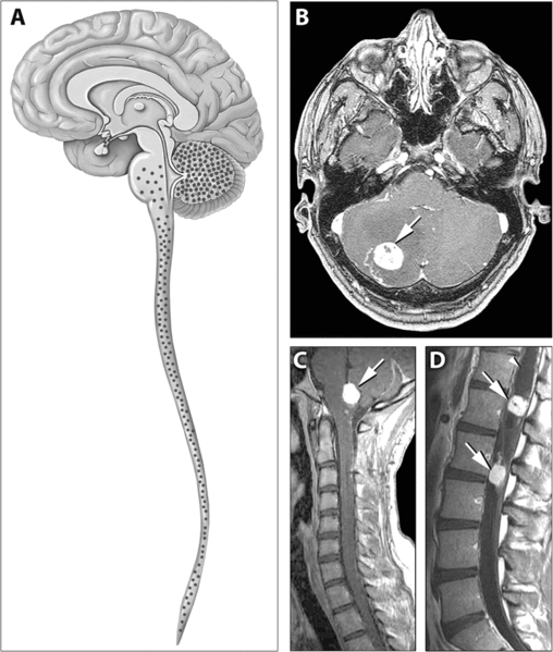
English: Distribution of Hemangioblastomas in the Central Nervous Systems of Study Patients
(A) Schematic representation of the distribution of CNS hemangioblastomas (red dots) in the 25 von Hippel-Lindau disease patients on MRI. Most (98%) of hemangioblastomas were found below the level of the tentorium in the cerebellum, brainstem, and spinal cord. (B–D) Contrast-enhanced MRI demonstrating representative locations of hemangioblastomas including the cerebellum (B), brainstem (C) and spinal cord (D). (B) Axial view through the cerebellum demonstrating a hyperintense enhancing hemangioblastoma (arrow) with surrounding edema (hypointense area surrounding the tumor) that frequently is associated with these lesions. (C) Sagittal view through the posterior fossa demonstrating a hyperintense enhancing brainstem (medullary) hemangioblastoma (arrow) with surrounding edema. (D) Sagittal view through the thoracic and lumbar spinal cord demonstrating two hyperintense enhancing hemangioblastomas (arrows). The superior tumor is associated with a large intraspinal cyst (syrinx) that is common with these neoplasms (arrowhead) |
| المصدر | http://medicine.plosjournals.org/perlserv/?request=get-document&doi=10.1371/journal.pmed.0040060 |
| المؤلف |
ترخيص
|
هذا الملف مُرخص تحت رخصة المشاع المبدع نسبة المصنف إلى مؤلفه 2.5 العامة
|
نُشِر هذا الملفُّ في دورية المكتبة العامَّة للعلوم. يُصرِّح الموقع الرسميِّ بأنَّ مُحتويات دوريات المكتبة العامَّة للعلوم مَنشورةٌ جميعها تحت رخصة المَشاع الإِبداعيِّ نسبة المُصنَّف إِلى مُؤَلِّفه (أو إلى إصداراتٍ سابِقة مِن الرُّخصة حسب تاريخ الإِصدار)، ما لم يذكر خلاف ذلك. |
تاريخ الملف
اضغط على زمن/تاريخ لرؤية الملف كما بدا في هذا الزمن.
| زمن/تاريخ | صورة مصغرة | الأبعاد | مستخدم | تعليق | |
|---|---|---|---|---|---|
| حالي | 13:43، 31 مايو 2007 | | 805 × 949 (278 كيلوبايت) | Filip em | Distribution of Hemangioblastomas in the Central Nervous Systems of Study Patients (A) Schematic representation of the distribution of CNS hemangioblastomas (red dots) in the 25 von Hippel-Lindau disease patients on MRI. Most (98%) of hemangioblastomas w |
استخدام الملف
الصفحة التالية تستخدم هذا الملف:
الاستخدام العالمي للملف
الويكيات الأخرى التالية تستخدم هذا الملف:
- الاستخدام في bs.wikipedia.org
- الاستخدام في ca.wikipedia.org
- الاستخدام في de.wikipedia.org
- الاستخدام في de.wikibooks.org
- الاستخدام في el.wikipedia.org
- الاستخدام في en.wikipedia.org
- الاستخدام في hy.wikipedia.org
- الاستخدام في it.wikipedia.org
- الاستخدام في ja.wikipedia.org
- الاستخدام في ru.wikipedia.org
- الاستخدام في sk.wikipedia.org
- الاستخدام في uz.wikipedia.org
- الاستخدام في zh.wikipedia.org